Watch: This amazing video shows Spina Bifida operation on unborn baby
A video made by Cleveland Clinic illustrating how it had successfully performed its first in utero foetal surgery to repair a spina bifida birth defect in a nearly 23-week-old foetus is now attracting significant attention on social media.
The clinic says that a team of clinicians, representing many specialties, performed the surgery in February, and the baby, a girl, was later delivered by caesarean section. Mother and daughter are doing well.

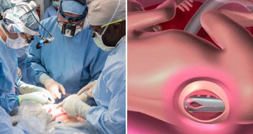
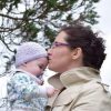
The video shows an overview of how the surgical team worked to repair the spina bifida lesion which had been observed on the baby’s spine. Surgeons carefully sew together several individual layers of tissue in order to cover the defect – all while the baby remains attached to the mother.
After the operation is complete, the womb is closed back up, and the baby continues in utero for the remainder of the pregnancy and is ultimately born by caesarean section. Although the surgery was performed in 2019, it seems to be attracting additional attention online now.
The surgical team, led by Darrell Cass, M.D., director of Fetal Surgery in Cleveland Clinic’s Fetal Center and a specialist who has performed more than 160 fetal surgeries since 2002, included Amanda Kalan, M.D., medical director of Cleveland Clinic’s Special Delivery Unit; Violette Recinos, M.D., and Kaine Onwuzulike, M.D., both pediatric neurosurgeons; Francine Erenberg, M.D., fetal cardiologist; and McCallum Hoyt, M.D. and Tara Hata, M.D., obstetric and pediatric anesthesiologists.
Spina bifida is a birth defect that is most often discovered during the routine anatomy scan typically performed when a fetus is around 18 weeks old.
The condition affects the lowest part of the spine and occurs when the neural tube does not fully close, causing the backbone that protects the spinal cord not to form as it should. This often results in damage to the spinal cord and nerves and can even lead to brain damage.
Spina bifida can affect a child’s lower leg strength and their ability to walk and run, as well as their ability to go to the bathroom and urinate adequately. According to the CDC, approximately 1,645 babies are born with spina bifida each year in the United States.
During the fetal repair surgery, a caesarean section-like incision is made and the mother’s uterus is exposed. An ultrasound is then used to locate the placenta and fetus.
The uterus is opened 4.5 cm and the back of the fetus is exposed, showing the spina bifida lesion. The surgeons then carefully suture several individual layers of tissue (myofascia, dura and skin) in order to cover the defect. After the uterus is closed back up, the fetus remains in the womb for the remainder of the pregnancy and is ultimately born by caesarean section.
“By successfully repairing the defect before birth, we’re allowing this child to have the best possible outcome and significantly improve her quality of life,” said Dr. Cass. “There are different measures of quality in determining success for fetal repairs and in this particular case, all metrics for maximum quality were achieved.”
The success of this surgery was based on two metrics – restoration of normal brain structure and the gestational age at birth.
Prior to fetal repair surgery, the back of the brain herniates down the spinal column – known as an Arnold-Chiari malformation – causing cerebrospinal fluid to back up and build pressure that can cause brain damage. Typically babies with spina bifida need shunts to decompress the built-up fluid after they are born.
When a successful fetal surgery repair is completed and the brain is examined one month later, the malformation is reversed and the back of the brain returns to a normal appearance, which was observed in this case.
The surgery was also successful by the second metric – when the baby is born. The average birth after fetal surgery repair is 34 weeks gestation. In this case, the baby was born at 36.5 weeks gestation – exceeding the average birth time by more than two weeks and giving the baby more time to develop and grow.
“Although the surgery was a success, spina bifida is never cured,” said Dr. Cass. “Moving forward, the baby will require ongoing supportive care provided by a multidisciplinary team of caregivers in our Spina Bifida Clinic, which will involve neurology, urology, orthopedics, developmental pediatrics and neurosurgery, among other specialists.”
The surgical team from Cleveland Clinic’s Fetal Center worked for more than a year to prepare for this first surgery, including making site visits to other centers, conducting simulations, and consulting with other experts in the field.
“I am honored to work with this amazing team of clinicians and to see our efforts come to fruition after preparing for so long,” said Dr. Cass, who joined Cleveland Clinic as director of Fetal Surgery in October 2017 to build its fetal surgery program after co-founding and co-directing Texas Children’s Fetal Center in Houston for 17 years. “Families in this region now have more options when it comes to making these types of decisions and we are thrilled to be able to provide the care needed for these complex cases.”
PHOTOS from the Cleveland Clinic show the skilled surgeons operating.

Darrell Cass, M.D., performs an in utero surgery to repair a spina bifida birth defect in a nearly 23-week-old fetus.


Pediatric neurosurgeons Violette Recinos, M.D. and Kaine Onwuzulike, M.D. perform repair
This article was first published in Gript and is printed here with permission
Featured
- Pathetic government backing SF to chase non-existent Repeal vote
- How did the Dáil vote on the Sinn Féin abortion bill?
- UK: Bill on babies born alive after an abortion receives First Reading in House of Lords
- WATCH: TD accuses SF of ignoring proof that 3-day wait saves lives
- NI: Woman cleared as abortion zone conviction collapses
- Witness at Cavan General Hospital
- Rally for Life 2026
- 109,000 payments to abortion providers in first 6 years
- HSE still has no guidelines for when babies survive abortions
- Family’s “unimaginable pain and grief” over murder of Natalie McNally and her unborn baby
- We remember 60,000 babies aborted on the 8th anniversary of Repeal
- DESPICABLE: YouTuber who aborted baby with Down Syndrome playing the victim after MASSIVE backlash
- ‘Pro-choice’ and sex-selective abortions
- ANALYSIS: What we learned from the debate on the SocDem’s abortion bill
- Wexford for Life Rally to take place on 20th June
- LOOK-BACK: How the fight to keep the 3 day wait won hearts and minds
- We Remember them 2026
- SHOCKING: 575 abortions carried out with no upper gestational age limit
-
 The Life Institute
-
 Ann Farmer
-
 Ann Marie Madden
-
 Baby Blogger
-
 Bernadette Smyth
-
 Caitlin Lawlor
-
 David Mullins
-
 Eadaoin R
-
 Elaine Cole
-
 Fionnuala Nic Mhathuna
-
 Guest Blogger
-
 Jennifer As
-
 Jennifer Fulwiler
-
 Jonathon Van Maren
-
 Kate Bryan
-
 Lydia Mead
-
 Maggie Walsh
-
 Mann Inside
-
 Maria Forrestal
-
 Maria Horan
-
 Marion Murphy
-
 Mary Anne
-
 Maurice O'Brien
-
 Megan Scallan
-
 Niamh Uí Bhriain
-
 Sandra Parda
-
 Sarah Howell
-
 Tim Jack